SolveItClass 9 · NCERT

NCERT Solutions · Class 9 Science Tissues in Action

25 questions · 25 still being checked

Revise, Reflect, Refine 3.1–3.10 (part 3 of 4)

  1. Exercise 3.1

    Meristematic tissues divide repeatedly. What property of their cells allows them to do this? (i) They have thick walls for protection. (ii) They contain large vacuoles that store nutrients. (iii) They have thin walls, dense cytoplasm and large prominent nucleus. (iv) They are functionally differentiated cells.

    Not cross-checked

    NCERT prints no numerical answer for this exercise, so this working has not been cross-checked against the book.

    NCERT’s answer
    (iii)
    (iii) They have thin walls, dense cytoplasm and large prominent nucleus.
    NCERT_Solution_Class9_Science_Ch3_RRR_Q3-1
    The chapter describes meristematic cells as small, with thin cell walls, a large and prominent nucleus, and dense cytoplasm containing many organelles — precisely the equipment for repeated division.
    The cells are also tightly packed with little or no intercellular space, and these features together are what allow continuous and rapid cell division.
    (i) is wrong — thick walls belong to sclerenchyma, whose cells are lignified and mostly dead, and dead cells do not divide.
    (ii) is wrong — vacuoles are generally absent in meristematic cells; a large vacuole would leave too little cytoplasm and too few organelles for division.
    (iv) is wrong — differentiation is what happens when a meristematic cell loses the ability to divide and turns into a permanent tissue; a differentiated cell is the opposite of a dividing one.
  2. Exercise 3.2

    If a plant is unable to transport food from leaves to roots which tissue is malfunctioning? (i) Xylem (ii) Phloem (iii) Epidermis (iv) Sclerenchyma

    Check this one against your book

    NCERT’s printed answer for this exercise does not match its own question. This working follows the question as printed.

    Phloem — option (ii).
    NCERT_Solution_Class9_Science_Ch3_RRR_Q3-2
    The chapter separates the two conducting tissues by cargo in its opening pages: "xylem transports water and minerals, while phloem transports food." Food travelling from leaves down to roots is therefore a phloem job.
    Section $\displaystyle 3.2.4$(iii), Conducting tissues, names the cells that do it. Phloem "consists of sieve tubes, companion cells, phloem parenchyma and phloem fibres", and "Sieve tubes transport food from leaves to other parts of the plant." The roots are among those "other parts", so a plant that cannot move food from leaves to roots has sieve tubes of the phloem that are not working.
    Why not (i) Xylem: the same section says xylem "transports water and minerals from the roots to other parts of the plant" — the wrong cargo, and the opposite direction to the one in the question.
    Why not (iii) Epidermis: the chapter files the epidermis under "(i) Protective tissue". It is "the outermost layer of the plant body", "a tightly packed, single layer of flat and rectangular cells" that "protects all parts of the plants". The jobs listed for it there are protection by the cuticle, absorption through root hair, and gaseous exchange and transpiration through stomata — never food transport. It is not one of the conducting tissues at all.
    Why not (iv) Sclerenchyma: it is listed as a simple permanent (supporting) tissue with "thick walls… due to deposition of lignin", of which "Most of these cells are dead". It supplies hardness and strength — coconut husk, walnut shell — not conduction.
    At a Glance states it once more: "Complex permanent tissues include xylem and phloem that transport water and food respectively to all parts of the plant."
    The book's printed Answer Key gives (iii) Epidermis, which does not agree with Section $\displaystyle 3.2.4$ or with the At a Glance summary. Both of those passages assign food transport from the leaves to the phloem's sieve tubes, and both classify the epidermis as a protective tissue with no conducting role, so the chapter's own text settles it the other way: the answer is (ii) Phloem, and the key entry should be treated as an error.
  3. Exercise 3.3

    Why are the epithelial tissues that line an animal’s internal organs usually only one or a few cells thick? (i) To store food efficiently. (ii) To provide maximum strength. (iii) To allow quick exchange of materials across them. (iv) To reduce friction.

    Check this one against your book

    NCERT’s printed answer for this exercise does not match its own question. This working follows the question as printed.

    To allow quick exchange of materials across them — option (iii).
    NCERT_Solution_Class9_Science_Ch3_RRR_Q3-3
    Table $\displaystyle 3.2$ pairs each epithelial structure with the function it serves, and its first row is exactly this case: function "Exchange: Helps in rapid diffusion of liquids and gases"; structure "Single layer of thin, flat cells"; location "Lining of the tissue in the blood vessels and lungs". A single thin layer keeps the diffusion path short, which is what makes the exchange rapid.
    The last row of the same table makes the same pairing for the gut: function "Absorption: Efficient uptake of nutrients, water, etc."; structure "Single layer of tall, pillar-like cells"; location "Lining of small intestine". Again a single layer wherever the tissue's job is moving material across itself.
    Section $\displaystyle 3.3.1$ gives the general statement: epithelial tissue "lines the internal organs, such as the mouth, lungs, blood vessels and intestine" and "helps in the absorption, secretion and movement of substances."
    Why not (ii) To provide maximum strength: Table $\displaystyle 3.2$ answers that with a different row. Where the body needs protection "from mechanical injury, friction and entry of microbes", the epithelium is "Many layers of cells; the outer cells are flat and tightly packed", located in "Skin, mouth and oesophagus". In this chapter strength comes from many layers — the opposite of the one-or-few-cell thickness the question asks about.
    Why not (iv) To reduce friction: friction appears in that same many-layered protection row, not in either of the thin, single-layer rows.
    Why not (i) To store food efficiently: storage is not among the five functions Table $\displaystyle 3.2$ lists for epithelium — they are exchange, protection, secretion, sensory functions and absorption.
    The book's printed Answer Key gives (ii) To provide maximum strength, which does not agree with Table 3.2. In that table maximum strength belongs to the "Many layers of cells" protective epithelium of skin, mouth and oesophagus, while the one-cell-thick lining of blood vessels and lungs is set against "rapid diffusion of liquids and gases". The chapter's own table therefore settles it the other way: the answer is (iii), and the key entry should be treated as an error.
  4. Exercise 3.4

    You can perform these two jumps (Fig. 3.21\displaystyle 3.21): Straight-leg jump — keep knees and ankles stiff. Normal jump — bend knees and ankles naturally. How did your ankle, knee and hip positions differ between the two jumps?NCERT_Question_Class9_Science_Ch3_RRR_Q3-4

    Not cross-checked

    NCERT prints no numerical answer for this exercise, so this working has not been cross-checked against the book.

    In the straight-leg jump all three joints stay nearly straight the whole time; in the normal jump all three bend first and then straighten together.
    Straight-leg jump. The knee stays locked straight before, during and after take-off, the hip stays extended with the trunk upright, and only the ankle can push. With one small joint doing all the work the jump is low, and on landing the stiff leg has nothing left to bend, so the shock passes straight up through the bones as a hard jolt.
    Normal jump — the crouch. The ankle bends forward, the knee bends, and the hip bends so the body leans slightly forward. All three joints are flexed at the same time.
    Normal jump — take-off. The three straighten in quick succession — hip, then knee, then ankle — each adding to the push, so the jump is far higher.
    Normal jump — landing. The sequence reverses: ankle, knee and hip bend again and absorb the shock gradually, so the landing is soft.
    Why the joints behave differently. The knee is a hinge joint and can only bend and straighten in one plane; the hip is a ball and socket joint, so it can also swing sideways and rotate as the body leans.
    What actually does the bending. The chapter is explicit that joints allow movement but cannot move the bones on their own — muscles contract and pull on the bones through tendons, and the movement appears at the joint.
  5. Exercise 3.5

    Which type of joint is involved when you bend your knees and ankles? (i) Ball and socket (ii) Hinge (iii) Pivot

    Check this one against your book

    NCERT’s printed answer for this exercise does not match its own question. This working follows the question as printed.

    Hinge joint — option (ii).
    NCERT_Solution_Class9_Science_Ch3_RRR_Q3-5
    Section $\displaystyle 3.5.2$ names the knee outright: "the elbow bends and straightens in one direction only like a door hinge… This type of joint is called a hinge joint. A similar hinge joint is present in the knee, where a small bone called the kneecap protects the joint." Bending your knee is precisely the movement that section is describing.
    Why not (iii) Pivot: Section $\displaystyle 3.5.3$ gives the pivot joint a different action in a different place — the skull is "connected to the backbone through a pivot joint, which allows the head to move side to side like a doorknob turning in its socket." That is a turn about an axis, not a bend, and it sits at the neck.
    Why not (i) Ball and socket: Section $\displaystyle 3.5.1$ places it at the shoulder, where "the rounded top of the upper arm bone fits into a shallow hollow of the shoulder bone", and the joint "allows forward, backwards, sideways and circular movements." The knee has no such freedom — recording that contrast is the point of Table $\displaystyle 3.5$, whose worked Elbow row reads Complete rotation: No, Partial rotation: No, Bending: Yes.
    On the ankle — this is reasoning, not chapter content. The chapter never classifies the ankle: it is not one of the body parts listed in Table $\displaystyle 3.5$ (Elbow, Shoulder, Knee, Neck, Fingers, Toes, Wrist), and none of Sections $\displaystyle 3.5.1$–$\displaystyle 3.5.4$ mentions it. Applying the chapter's own definition, pointing the toes down and up is bending in one direction only rather than rotating, which fits the hinge description and not the pivot's side-to-side turn. Treat that as an inference from the chapter's criteria, not as a statement the book makes.
    The book's printed Answer Key gives (iii) Pivot, which does not agree with Sections $\displaystyle 3.5.2$ and 3.5.3. Section $\displaystyle 3.5.2$ says in so many words that a hinge joint "is present in the knee", and Section $\displaystyle 3.5.3$ assigns the pivot joint to the skull-and-backbone connection at the neck, so the chapter's own text settles it the other way: the answer is (ii) Hinge, and the key entry should be treated as an error.
  6. Exercise 3.6

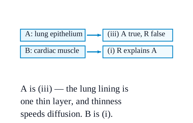
    In each of the following cases (A, B, C and D), choose the correct option as given below: (i) Both (A) and (R) are true, and (R) is the correct explanation of (A). (ii) Both (A) and (R) are true, but (R) is not the correct explanation of (A). (iii) (A) is true, but (R) is false. (iv) (A) is false, but (R) is true. A. Assertion: Epithelium is well-suited for gas exchange in the lungs. Reason: It consists of multiple layers of tall cells that slow down diffusion. B. Assertion: Cardiac muscle can contract continuously without fatigue. Reason: Cardiac muscle cells have a high number of mitochondria and an abundant blood supply.

    Not cross-checked

    NCERT prints no numerical answer for this exercise, so this working has not been cross-checked against the book.

    NCERT’s answer
    A (iii) B (i) C (iv) D (iii)
    A — (iii): (A) is true, but (R) is false.
    NCERT_Solution_Class9_Science_Ch3_RRR_Q3-6
    Assertion true: Table $\displaystyle 3.2$ places a single layer of thin, flat cells in the lining of the lungs precisely because it helps rapid diffusion of liquids and gases.
    Reason false on two counts: that lining is one cell thick, not multiple layers of tall cells; and its thinness speeds diffusion rather than slowing it. Multiple layers of tall cells describe the protective and absorptive epithelia instead.
    B — (i): both true, and (R) is the correct explanation of (A).
    Assertion true: the chapter says cardiac muscles work tirelessly and rhythmically, enabling the heart to beat throughout life without fatigue.
    Reason true and genuinely explanatory: many mitochondria mean a steady supply of energy, and an abundant blood supply keeps delivering the oxygen and nutrients needed to make it — so the muscle never runs an energy debt, which is exactly why it does not tire.
    C — (iv): (A) is false, but (R) is true.
    Assertion false: tendons connect muscle to bone; it is ligaments that connect bone to bone, and Table $\displaystyle 3.4$ sets the two out side by side.
    Reason true, and it is the chapter's own sentence: when a muscle contracts, the tendon transmits this force to the bone, resulting in movement at a joint. Note that the reason quietly states the correct fact that the assertion gets wrong.
    D — (iii): (A) is true, but (R) is false.
    Assertion true: §$\displaystyle 3.5.2$ says a hinge joint bends and straightens in one direction only, like a door hinge — the elbow and the knee.
    Reason false: bone ends shaped to allow sliding in all directions would give a ball and socket joint. The hinge shape is what restricts movement to one plane, so the reason describes the opposite of what a hinge does.
  7. Exercise 3.7

    Plot a graph between the age of a tree (in years) on the x-axis and the diameter of the tree (in cm) along with the number of annual rings formed over time on the y-axis, using the data given in the Table 3.7. increase in the diameter of stem and number of annual rings S. No. 1. 2. 3. 4. 5. 6. (i) Analyse the graph in terms of the diameter of the stem over time and share the interpretation. (ii) What is the relation between the diameter of the teak tree to the annual rings formed? (iii) Which specialised tissue is responsible for the girth of the stem and where is it located?

    Not cross-checked

    NCERT prints no numerical answer for this exercise, so this working has not been cross-checked against the book.

    The graph to draw. One graph carrying two plotted lines against the same axes.
    x-axis: Age of the teak tree (years), scaled \(\displaystyle 0 \) to \(\displaystyle 40 \) in steps of \(\displaystyle 5 \).
    y-axis: Diameter (cm) and Number of annual rings, scaled \(\displaystyle 0 \) to \(\displaystyle 40 \) in steps of \(\displaystyle 5 \) — both quantities happen to share the same range, so one scale serves both.
    Line $\displaystyle 1$ — DBH (cm): plot \(\displaystyle (5,4),\ (10,8),\ (20,24),\ (25,28),\ (30,32),\ (40,40) \) and join the points.
    Line $\displaystyle 2$ — Number of annual rings: plot \(\displaystyle (5,5),\ (10,10),\ (20,20),\ (25,25),\ (30,30),\ (40,40) \) and join the points. This comes out as a perfectly straight line through the origin at \(\displaystyle 45^\circ \).
    Mark each plotted point clearly, use a different symbol or line style for the two lines, and add a key naming them.
    (i) Interpretation of the diameter curve
    The diameter increases steadily throughout — the teak tree never stops thickening, because its lateral meristem keeps dividing year after year.
    Average rate over the whole record: \(\displaystyle \dfrac{40 - 4}{40 - 5} = \dfrac{36}{35} \approx 1.03 \) cm per year.
    The rate is not uniform, interval by interval:
    \(\displaystyle 5 \to 10 \) yr: \(\displaystyle \dfrac{8-4}{5} = 0.8 \) cm/yr
    \(\displaystyle 10 \to 20 \) yr: \(\displaystyle \dfrac{24-8}{10} = 1.6 \) cm/yr
    \(\displaystyle 20 \to 25 \) yr: \(\displaystyle \dfrac{28-24}{5} = 0.8 \) cm/yr
    \(\displaystyle 25 \to 30 \) yr: \(\displaystyle \dfrac{32-28}{5} = 0.8 \) cm/yr
    \(\displaystyle 30 \to 40 \) yr: \(\displaystyle \dfrac{40-32}{10} = 0.8 \) cm/yr
    So the curve is steepest between years \(\displaystyle 10 \) and \(\displaystyle 20 \), where girth grows at twice the rate of every other stretch, and settles back to a steady \(\displaystyle 0.8 \) cm/yr after that.
    This matches the chapter's account of annual rings: some are wide and some narrow, reflecting favourable or unfavourable growth conditions in that year. The years \(\displaystyle 10\!-\!20 \) were the favourable ones for this tree.
    (ii) Relation between diameter and annual rings
    The number of annual rings equals the age in years exactly — \(\displaystyle 5, 10, 20, 25, 30, 40 \) rings at \(\displaystyle 5, 10, 20, 25, 30, 40 \) years — so one ring is laid down each year, and counting rings gives the tree's age.
    Diameter and ring count rise together: the more rings, the greater the diameter. Each ring is one year's fresh layer of cells added to the girth.
    The relation is close to proportional but not exactly so, because rings differ in width: \(\displaystyle \dfrac{4}{5} = 0.8 \) cm per ring at age \(\displaystyle 5 \), \(\displaystyle \dfrac{24}{20} = 1.2 \) cm per ring at age \(\displaystyle 20 \), and \(\displaystyle \dfrac{40}{40} = 1.0 \) cm per ring at age \(\displaystyle 40 \).
    Practical use: from a cut trunk, ring count gives the age and ring widths give the climate record of the years the tree grew through.
    (iii) The tissue responsible for girth
    The lateral meristem.
    It is a meristematic tissue of actively dividing cells arranged in a ring along the circumference of the stem — visible in the T.S. of the sunflower stem in Fig. $\displaystyle 3.7$, lying between the phloem outside and the xylem inside.
    It divides and produces new cells both inside and outside in concentric layers, and that is what increases the diameter of the stem.
    NCERT_Solution_Class9_Science_Ch3_RRR_Q3-7
  8. Exercise 3.8

    In a forest, it was observed that one of the trees was severely debarked by an elephant to meet its food requirements, as the bark is a rich source of nutrients (Fig. 3.22\displaystyle 3.22). Based on your learning, answer the following: (i) Which function(s) of the tree is/are hampered by debarking? (ii) Which plant tissue would be affected by further damage to the tree trunk even after debarking? (iii) Which function of the tree would be hampered if the tissues beneath the bark were severely damaged? (iv) What assumptions are you making to answer the questions above? How would the answer change if your assumptions are also changed?NCERT_Question_Class9_Science_Ch3_RRR_Q3-8

    Not cross-checked

    NCERT prints no numerical answer for this exercise, so this working has not been cross-checked against the book.

    (i) Functions hampered by debarking
    Protection and food transport are the two that fail first.
    The bark is made of cork cells — dead, compactly arranged, and containing a substance that makes them impermeable to water and gases. Strip it and the trunk loses its shield against water loss, mechanical injury, harmful microorganisms and extreme environmental conditions.
    Immediately beneath the bark lies the phloem. If it is torn away with the bark, food made in the leaves can no longer be carried down to the stem and the roots, and the roots begin to starve because they cannot photosynthesise for themselves.
    The exposed wound is an open door for parasites and pathogens — the chapter notes that the cuticle and outer covering exist partly to prevent invasion by parasites.
    (ii) Tissue affected by further damage to the trunk
    Working inwards from the surface, the order in Fig. $\displaystyle 3.7$ is epidermis/cork → ground tissue → phloemlateral meristemxylem.
    So the phloem is hit next, and then the lateral meristem (cambium) just inside it. Deeper damage reaches the xylem.
    (iii) Function hampered if the tissues beneath the bark are severely damaged
    Loss of phloem → transport of food from leaves to stem and roots stops completely; the roots starve.
    Loss of lateral meristem → the tree can no longer grow in girth, cannot lay down new phloem, new xylem or new cork, and so cannot heal the wound or add another annual ring.
    Loss of xylem → transport of water and minerals from roots to leaves stops, and the tree also loses the mechanical strength xylem provides. The leaves wilt, photosynthesis fails, and the tree dies.
    (iv) Assumptions, and how the answers would change
    Assumed that the tree is a dicot with secondary growth, so it has bark, a cambium ring and annual rings at all.
    Assumed that "bark" here means the dead cork layer together with the phloem just under it — that is, the elephant took conducting tissue and not merely the dead covering.
    Assumed that the damage runs all the way round the trunk, not just across a patch.
    Assumed the cambium underneath is still alive.
    If only the dead cork was scraped off and phloem plus cambium survive, the tree loses protection alone. The cork cambium can divide and make fresh cork, and the tree recovers with a scar.
    If the damage is a patch rather than a complete ring, food can still travel down through the intact phloem on the undamaged side, and the tree survives.
    If it is a complete ring and the phloem is gone all round, the roots are permanently cut off from food. They starve and die, and the whole tree follows — even though the xylem is intact and the leaves stay green for a while, because water can still go up while food cannot come down.
  9. Exercise 3.9

    Aamrapali observed that a young mango sapling’s stem bends flexibly during monsoon winds and does not break. Which tissue is responsible for this flexibility? Predict and provide your explanation of the impact if the existing tissue was replaced by sclerenchyma.

    Not cross-checked

    NCERT prints no numerical answer for this exercise, so this working has not been cross-checked against the book.

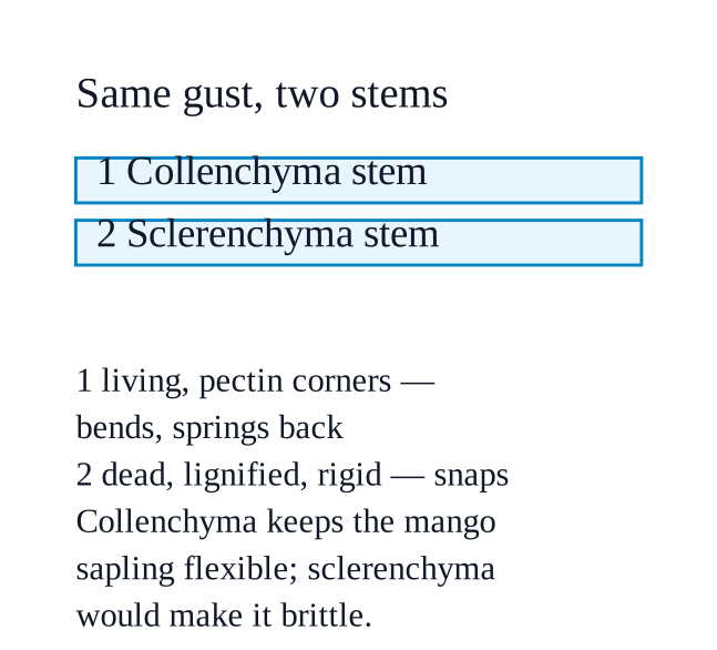
    Collenchyma is the tissue responsible for the flexibility of the mango sapling's stem.
    NCERT_Solution_Class9_Science_Ch3_RRR_Q3-9
    Its cells are living, with their corners unevenly thickened by pectin — a chemical the chapter compares to rubber for the flexibility it gives.
    That combination supports the stem without making it rigid, which is precisely what the chapter credits collenchyma with: it allows parts of the plant like stems and tendrils to bend without breaking.
    In a monsoon gust the stem gives way, lets the wind pass, and springs back.
    If sclerenchyma replaced it, the stem would become stiff and brittle and would snap.
    Sclerenchyma cells have thick walls due to deposition of lignin, making them hard and strong and forming the woody structure — and most of these cells are dead.
    Hard is not the same as tough: a rigid rod cannot absorb the energy of a gust the way a flexible one can, so the force would go into breaking the stem instead of bending it.
    Being dead and lignified, sclerenchyma also cannot stretch or elongate, so a young growing stem would lose the ability to extend and to recover its shape after being bent.
    The chapter's own contrast makes the point in two familiar objects: coconut husk fibre (sclerenchyma) is hard and brittle, while a coriander leaf stalk (collenchyma) is soft and flexible. A sapling built like coconut husk would not survive a monsoon.
  10. Exercise 3.10

    Sohan designed an experiment for the regeneration of sugarcane, where he used cuttings to grow sugarcane. He used two types of cuttings, type ‘A’ and type ‘B’ (Fig. 3.23\displaystyle 3.23). After a few weeks, type ‘B’ cuttings sprouted and developed into sugarcane plants, whereas the type ‘A’ cuttings did not sprout. (i) Why were the type ‘B’ cuttings able to grow as sugarcane but type ‘A’ could not? (ii) What difference was present in type ‘B’ compared to type ‘A’? (iii) What observation or measurement was made to determine whether this change had an effect? (iv) What parameters should be kept the same for both types of cuttings to ensure a fair comparison?NCERT_Question_Class9_Science_Ch3_RRR_Q3-10

    Not cross-checked

    NCERT prints no numerical answer for this exercise, so this working has not been cross-checked against the book.

    (i) Why type B grew and type A did not
    Type B contained a node; type A did not.
    A node is the point on the stem where branches or leaves arise, and it carries a bud together with intercalary meristem, which the chapter locates at the base of the internode or just above the node.
    Meristematic cells divide continuously, so the bud at the node can sprout into a new shoot, after which the cutting puts out roots and grows into a whole sugarcane plant.
    Type A was cut from the internode — the part of the stem between two nodes. Its cells are permanent tissue: through differentiation they have lost the ability to divide, and a tissue that cannot divide cannot produce a new shoot.
    This is the same principle as the hedge that grows bushy after trimming and the grass that grows back after mowing — the intercalary meristem at the nodes is what regenerates the plant.
    (ii) The difference present in type B
    The presence of at least one node, with its bud and its intercalary meristematic tissue.
    Type A had only internode tissue, entirely permanent and non-dividing.
    (iii) Observations and measurements made
    Whether each cutting sprouted at all — a simple yes/no for every cutting.
    The number of cuttings that sprouted out of the total in each group, e.g. \(\displaystyle \dfrac{8}{10} \) of type B against \(\displaystyle \dfrac{0}{10} \) of type A.
    The number of days taken to sprout.
    The number of shoots produced per cutting.
    The length of each new shoot in cm, measured at fixed intervals over the few weeks.
    Whether roots formed, and how many.
    (iv) Parameters to keep the same for a fair comparison
    Everything except the one factor being tested — the presence of a node.
    Same sugarcane variety, and cuttings taken from the same parent plant of the same age.
    Same length and thickness of every cutting.
    Equal numbers of cuttings in group A and group B.
    Same soil or growing medium, same pot size, and the same depth and orientation of planting.
    Same watering schedule, light, temperature and humidity for both groups.
    Same period of observation, with both groups checked on the same days.