SolveItClass 9 · NCERT

NCERT Solutions · Class 9 Science Cell: The Building Block of Life

24 questions · 24 still being checked

Pause and Ponder 2.1–2.8 (part 1 of 3)

  1. Exercise 2.1

    What argument would you give for the necessity of a cell wall in plants usually fixed in one place versus in animals usually moving from one place to the other?

    Not cross-checked

    NCERT prints no numerical answer for this exercise, so this working has not been cross-checked against the book.

    A cell wall is a support structure, and a plant that cannot walk away from trouble needs support far more than an animal does.
    A plant is fixed in one place, so it cannot escape wind, rain or heat — it must stand up to them where it stands. The rigid cell wall gives each of its cells the strength to do that.
    The wall also keeps leaves and flowers firm, holds their shape, and helps the whole plant stay upright.
    That rigidity costs the plant nothing it needs, because the cell wall is permeable: water and dissolved minerals still pass straight through it, so roots go on absorbing water and nutrients from the soil.
    The wall is what saves a plant cell in a concentrated solution — the contents shrink away inside, but the cell keeps its box shape (Fig. $\displaystyle 2.9$).
    An animal moves, so its cells must bend, stretch and change shape. Animal cells have no cell wall, and this flexibility supports the movement and functioning of animal tissues.
    A wall around animal cells would lock every tissue rigid, and the animal could not move at all.
    NCERT_Solution_Class9_Science_Ch2_PP_Q2-1
  2. Exercise 2.2

    What consequences would you predict for a plant cell if its cell wall were to become as flexible as a cell membrane?

    Not cross-checked

    NCERT prints no numerical answer for this exercise, so this working has not been cross-checked against the book.

    The cell would lose its shape and its firmness, and the plant built from such cells would no longer stand up.
    In a concentrated sugar or salt solution the whole cell would collapse inwards, like a cheek cell, instead of only its contents pulling away while the outline held (compare Fig. 2.9a and b).
    In plain water the opposite failure: water would keep entering by osmosis with nothing rigid to push back, so the cell would swell and could burst.
    Cells could no longer press firmly against one another, so stems would droop and leaves and flowers would go limp even when the plant had plenty of water.
    The plant would have no defence left against wind and rain — which is exactly the stress the cell wall exists to resist.
    Water and minerals would still get in, since the wall was already permeable; what would be lost is only the rigidity.
    NCERT_Solution_Class9_Science_Ch2_PP_Q2-2
  3. Exercise 2.3

    Why is it important to cut the two potato pieces in roughly equal size and measure their initial weight before placing them in different liquids? membrane, In addition to the nucleus, the cytoplasm contains several sub-cellular Vacuole Chloroplast Cell wall Ribosome cell (Fig. 2.10a, b and c). A bacterial cell lacks a well-defined nucleus and membrane-boundNCERT_Question_Class9_Science_Ch2_PP_Q2-3

    Not cross-checked

    NCERT prints no numerical answer for this exercise, so this working has not been cross-checked against the book.

    Because the two pieces must differ in only one thing — the liquid they are placed in.
    Roughly equal size means roughly equal surface area of cell membrane exposed to the liquid, so water enters or leaves at comparable rates. A large piece and a small piece would change by different amounts for reasons that have nothing to do with the solution.
    The initial weight is the baseline. The whole result of the activity is the change, \(\displaystyle \text{change} = \text{final weight} - \text{initial weight} \), and with no initial value there is no change to calculate.
    It turns "the potato looks a little swollen" into a measured number, so the gain in Beaker A and the loss in Beaker B can be shown rather than claimed.
    This is what makes it a fair test: one variable changed, everything else held the same.
  4. Exercise 2.4

    material enclosed by a membrane) Primitive nucleus or nucleoid (genetic

    Not cross-checked

    NCERT prints no numerical answer for this exercise, so this working has not been cross-checked against the book.

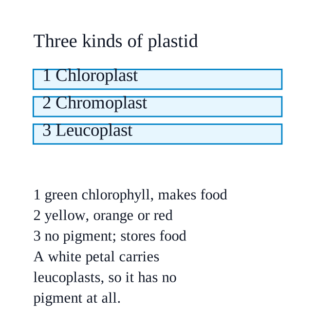
    No — a white petal contains no pigment at all.
    NCERT_Solution_Class9_Science_Ch2_PP_Q2-4
    Its plastids are leucoplasts, the plastids that lack pigments and are therefore colourless; their job is storing food such as starch, oils or proteins.
    Colour in petals and fruits comes from chromoplasts, whose pigments are yellow, orange or red; green comes from chlorophyll inside chloroplasts.
    With no pigment to absorb any part of the light falling on it, the petal throws nearly all of that light back, and what reaches our eyes is white.
    The chapter's point about bright colours also fits: pigments exist to attract pollinators and fruit-eating animals, so a flower that attracts by scent or shape has no need to make one.
  5. Exercise 2.5

    material without membrane around it)

    Not cross-checked

    NCERT prints no numerical answer for this exercise, so this working has not been cross-checked against the book.

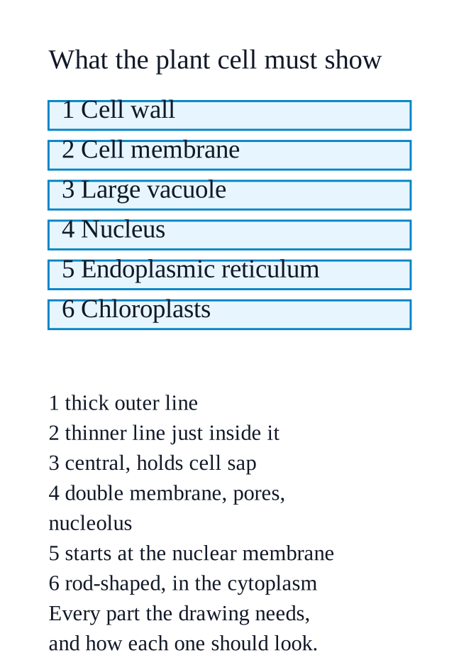
    Draw the plant cell — it is the one that shows every clue, including the rod-shaped chloroplasts.
    NCERT_Solution_Class9_Science_Ch2_PP_Q2-5
    Outline, two lines and not one: a thick outer rectangle with rounded corners for the cell wall, and a thinner line drawn just inside it, following it the whole way round, for the cell membrane.
    Vacuole: one large central space filling most of the cell, bounded by a single line and labelled vacuole (cell sap). Everything else goes in the narrow band of cytoplasm between it and the membrane.
    Nucleus (clue i): a dark round body pushed to one side. Draw its boundary as two closely spaced lines — the double-layered nuclear membrane — leave two or three gaps in it for the nuclear pores, and put a small solid circle inside for the nucleolus.
    Endoplasmic reticulum (clue ii): begin it at the nuclear membrane, so it clearly runs out of the nuclear envelope, and let it spread through the cytoplasm as a branching network of long flattened tubes. Dot one stretch with tiny beads and label it rough ER (ribosomes attached); leave another stretch undotted and label it smooth ER.
    Mitochondria (clue iii): two or three rod-shaped bodies, each with a smooth outer line and an inner line thrown into finger-like folds — label those folds cristae.
    Chloroplasts (clue iii): three or four rod-shaped bodies with a double boundary, a few small discs drawn inside them, and the space around the discs labelled stroma.
    Golgi apparatus: a stack of four or five flattened, slightly curved sacs, with tiny circles budding off the ends as vesicles.
    Ribosomes: scatter a few free dots in the cytoplasm as well as the ones sitting on the rough ER.
    Label the remaining space cytoplasm, run a ruled label line from every single part to its name written outside the cell, and title the figure "A typical plant cell".
    If you pick the animal cell instead: no cell wall and no chloroplasts, an irregular rounded outline, only small scattered vacuoles, and add lysosomes as small single-membrane sacs.
  6. Exercise 2.6

    Membrane-bound organelles Ready to Go Beyond In eukaryotic cells, a network of fine fibres forms the cytoskeleton, which provides structural support, maintains cell shape, and enables cell movement and internal transport. It is visible only under an electron microscope as a separate entity. The cytoplasm may also store starch (in plant cells), or crystals of calcium oxalate or silica (in some plant cells). These are known as cell inclusions.

    Not cross-checked

    NCERT prints no numerical answer for this exercise, so this working has not been cross-checked against the book.

    Because surface area is what a mitochondrion works with, and many small ones carry far more of it than one giant one of the same volume.
    Cellular respiration happens on the inner membrane. That is precisely why the inner membrane is thrown into cristae, finger-like projections that increase the surface area for the chemical reactions and so make more energy available. More membrane means more sites where ATP can be made.
    Surface area grows as the square of the size while volume grows as the cube, so anything larger has less surface per unit of volume:
    one cube of side \(\displaystyle 2 \): surface \(\displaystyle = 6 \times 2^2 = 24 \), volume \(\displaystyle = 2^3 = 8 \), ratio \(\displaystyle = 24 / 8 = 3 \)
    eight cubes of side \(\displaystyle 1 \): surface \(\displaystyle = 8 \times 6 \times 1^2 = 48 \), volume \(\displaystyle = 8 \times 1 = 8 \), ratio \(\displaystyle = 48 / 8 = 6 \)
    the same total volume, but twice the working surface once it is split up.
    Small mitochondria can also be spread right through the cytoplasm and sit next to whatever needs ATP; one giant mitochondrion would leave the far corners of the cell short.
    And a cell can build, repair or dispose of them one at a time as its energy demand changes — losing one of many is not losing everything.
    Also in the supplied text — Activity $\displaystyle 2.4$, Table $\displaystyle 2.1$
    Cell structureBacterial cellPlant cellAnimal cell
    Cell membranePresentPresentPresent
    Cell wallPresentPresentAbsent
    CytoplasmPresentPresentPresent
    Well-defined nucleusAbsentPresentPresent
    Primitive nucleus (nucleoid)PresentAbsentAbsent
    Membrane-bound organellesAbsentPresentPresent
    Also in the supplied text — "Which cells in Fig. $\displaystyle 2.10$ are prokaryotic and eukaryotic?"
    Fig. $\displaystyle 2.10$(a), the bacterial cell, is prokaryotic — no well-defined nucleus (a nucleoid instead) and no membrane-bound organelles.
    Fig. $\displaystyle 2.10$(b) the plant cell and Fig. $\displaystyle 2.10$(c) the animal cell are eukaryotic — each has a membrane-bound nucleus and several membrane-bound organelles.
    NCERT_Solution_Class9_Science_Ch2_PP_Q2-6
  7. Exercise 2.7

    Cut the tip portion of the root on the slide and put a coverslip. Gently squash the coverslip with your thumb to spread the cells on the slide.

    Not cross-checked

    NCERT prints no numerical answer for this exercise, so this working has not been cross-checked against the book.

    The cut would not heal.
    NCERT_Solution_Class9_Science_Ch2_PP_Q2-7
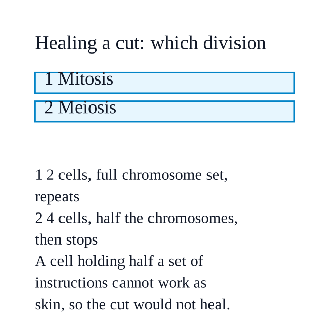
    Skin repairs itself by mitosis, the division that gives two daughter cells with the same DNA and the same number of chromosomes as the parent — so the new skin is an exact replacement for the skin that was lost.
    Meiosis halves the chromosome number. Each new cell would carry only half the DNA of the skin cell it came from, and a cell holding half a set of instructions cannot work as a skin cell.
    Meiosis also stops after making four cells; its products are gametes, not cells that go on dividing. The wound would get one round of unusable cells and then nothing, whereas mitosis repeats again and again until the gap is filled.
    Meiosis deliberately creates variation, so even the few cells formed would differ genetically from the skin around them instead of matching it.
    On top of all this the body would be making gamete-like cells in entirely the wrong place: the chapter is explicit that meiosis occurs only in the cells of reproductive organs — testes and ovaries.
  8. Exercise 2.8

    Observe the slide under a microscope.

    Not cross-checked

    NCERT prints no numerical answer for this exercise, so this working has not been cross-checked against the book.

    Yes, the squashed and stained root tip shows the cells clearly — and they are not all alike.
    Aceto-carmine stains the genetic material, so in some cells you see an ordinary nucleus, in others darkly stained thread-like or rod-shaped bodies instead, and in a few the stained material appears drawn towards opposite ends of the cell.
    Why they differ: the cells at a growing root tip divide continuously, so at the moment the root was fixed in aceto-alcohol every cell had been caught at a different point in cell division. The slide is a set of snapshots of different stages, which is exactly what Fig. $\displaystyle 2.17$ shows.
    Which stage comes first cannot be settled from this chapter — it names neither the stages nor their order, and says the stages of cell division are taken up in higher grades. All that can be said here is that the differences are stages of one continuous process, not different kinds of cell.
    NCERT_Solution_Class9_Science_Ch2_PP_Q2-8